
More Clearly
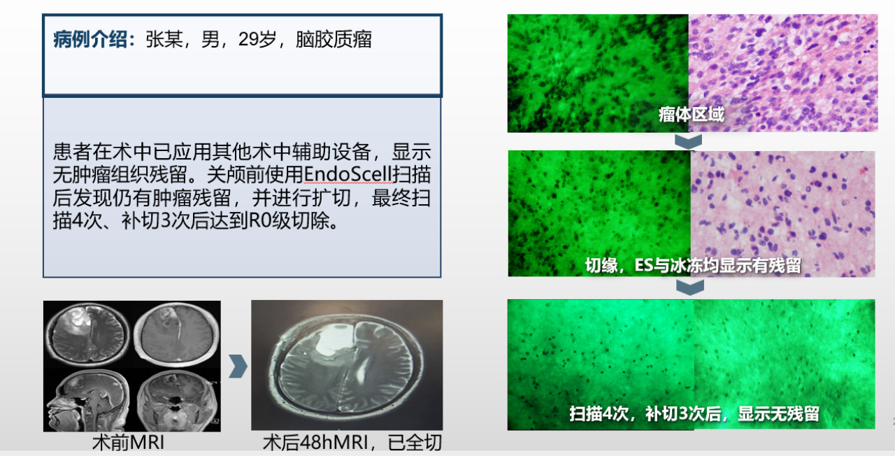
Intraoperative imaging technologies like ultrasound, MRI, fluorescence, and navigation often struggle to accurately locate tumor margins. EndoSCell?, with 1,400X magnification, extends the surgeon's view from surface tissues to the cellular level, allowing for direct visualization of tumor cells.

Faster
Traditional intraoperative frozen section pathology takes about 30 minutes for results, requiring multiple pathology reviews and resections. With EndoSCell?, whole-tumor cavity staining is achieved in 2 minutes, and a complete scan is done in 10-15 minutes. The imaging accuracy and sensitivity are comparable to cell smear slides, making margin assessments faster and more efficient.

Comprehensive
EndoSCell? uses Roving Scan to provide comprehensive coverage of the tumor resection area, enabling a full cavity search without multiple pathology reviews. The ESI imaging method easily differentiates cells in various conditions, ensuring no cancer cells are missed.

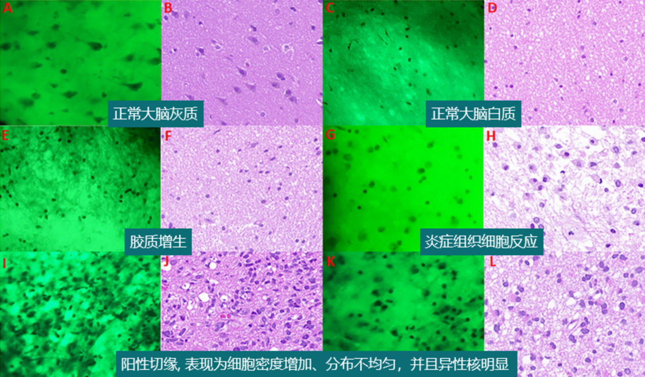
EndoScell鏡頭下的細胞組織與H&E stain結果對比
Courtesy of Dr. Xiang Zou, Huashan Hospital. Including unpublished data.